In-house Imaging Services for Injuries in Pittsburgh, PA
LineUp Scans For Lower & Upper Extremity
At Prisk Orthopaedics and Wellness in Pittsburgh, PA, we revolutionize orthopedic imaging with LineUP bilateral, weight-bearing CT technology. Our cutting-edge in-house imaging services offer unparalleled clarity up to the knee and cover essential areas like the hand, wrist, and elbow. Say goodbye to traditional imaging limitations and experience a new dimension of precision and accuracy in diagnosing orthopedic conditions.
We invite sports players, dancers, and anyone suffering from foot and ankle problems to contact us for a consultation and testing. To receive your evaluation and begin discussing your care needs, please contact Dr. Prisk at (412) 525-7692 or submit an online appointment request form.
Frequently Asked Questions
What are the benefits of using the LineUP CT scan?
The LineUP CT scan offers numerous benefits compared to traditional imaging methods. It provides a higher level of detail, allowing for better visualization and detection of small abnormalities. The 3D images also allow for more precise measurements and planning for surgical procedures. Additionally, the LineUP CT scan uses a lower dose of radiation than traditional CT scans, making it safer for patients.
The LineUP CT scan is suitable for anyone experiencing orthopedic issues in areas such as the

- Pre-operative planning
- Post-operative planning
- Diagnosis of fractures
- Evaluation of arthritic joints, bunion deformity, ankle instability, foot alignment, and other foot and ankle concerns
- Evaluation of hand, wrist, and elbow concerns
- Sesamoid position and condition (i.e. cases of a bone embedded in a tendon)
How Exactly Does The Computed Tomography Scan Work?

The LineUP CT scan works by using a series of X-ray images taken from different angles around the body to create detailed cross-sectional images. These images are then reconstructed by a computer to provide a 3D view of the area being scanned. This allows for a more accurate and comprehensive evaluation of orthopedic conditions.
Rather than limiting doctors to working from the “single angle” of traditional imaging, CT technology allows doctors to see every aspect of a patient’s foot and ankle health. And by allowing patients to receive standing (weight-bearing) imaging tests, patients can receive the most accurate assessment of the functional capabilities of their afflicted foot and ankle.
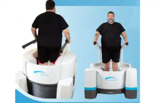
How Are LineUP's Standing CT Scans Taken?
To get a standing CT scan, you step on to a circular platform. The platform is about 2 inches off the ground, and there are handlebars on either side of the platform to assist you. There is a cushioned seat attached to the back of the system in case you are unable to stand. You stand in place as the entry doors, which are not taller than the height of an average person’s knee, close in front of you. The operator will start the scan, and you will be asked to stay completely still for 30 – 60 seconds. Then the doors will re-open and you will be able to step out.
How Is This Standing CT Scan Different From A Traditional CT Scan?
Traditional computed tomography requires patients to lie still on a sliding table. As a result, the foot is in a relaxed position. Traditional imaging must also make several rotations around the patient to create images of a particular pain point.
But when a scan is taken in the standing position, physicians can evaluate the true alignment of a person’s bones and joints. For example, physicians can clearly see if the ankle is rotated out of position, or if joint space is compromised in the midfoot. A standing CT also uses cone beam CT technology, which means the X-Ray tube only needs to make one revolution around the foot and ankle to create images of a particular pain point.
Although there are methods to “simulate” the standing position while the patient is lying on a table, the best way to evaluate the state of a foot and ankle problem is to have the patient stand.
What Do I Need To Know About Radiation Doses?
A standing CT scan exposes you to about 2 – 6 micro Sieverts of radiation. To put this in perspective, the average American is exposed to about 8 micro Sieverts of radiation a day from his or her environment. (A passenger on a flight from Los Angeles to New York is exposed to about 40 micro Sieverts of radiation.)
Peer-reviewed studies state the radiation dose of a standing CT scan is insignificant and should not be a deciding factor when determining if a patient needs a scan.
How Is My Scan Used?
After image testing is completed, your treating physician and a radiologist typically work together to analyze your scan. You may be provided with a radiology report, and you can request a copy of your scan. It will eventually also be provided to you as a DICOM (Digital Imaging and Communications in Medicine) file. There are a number of free DICOM viewers available for download if you would like to view your scan.
Give us a call at (412) 525-7692 or submit an online appointment request form, if you have any questions.