
High ankle sprains are different from the typical “rolled ankle.” Instead of the outer ligaments alone, a high ankle sprain injures the syndesmosis, the group of ligaments that connect the tibia and fibula just above the ankle joint. When these tissues are stretched or torn, the bones can spread apart under load, creating ankle instability, pain with pivoting, and delayed return to work or sport. Because the problem sits deeper and higher than a standard sprain, it is easier to miss and more likely to become chronic if not handled correctly.
What Happens?
A classic mechanism is a twist with the foot planted, often with external rotation. Pain sits above the ankle joint line and worsens when you push off, climb stairs, or pivot. Swelling can be modest compared with the pain. Walking on uneven ground or stepping out of a work vehicle can feel unstable or sharp.
How is it Diagnosed?
A detailed exam looks for tenderness along the syndesmosis, pain with external rotation stress, and signs of medial pain that can suggest deltoid ligament involvement. I start with weight‑bearing ankle radiographs to assess alignment. When I need more detail about subtle widening or rotational malalignment under load, I use weight‑bearing CT at our office in Monroeville. MRI helps evaluate the ligaments and can screen for cartilage damage or bone bruising but may underestimate instability because it is done without load. Taken together, these tools let me classify the injury as stable or unstable and decide on the safest path to recovery.
WHY SURGERY? When does it make sense?
Stable injuries can often be treated without surgery using a boot, protected weight bearing, and a progressive rehab plan. Surgery becomes the better option when there is clear instability, associated fractures, failure of non‑operative care, or persistent symptoms that keep you from your job or sport. My goals in surgery are simple: restore the normal relationship of the tibia and fibula, treat any cartilage or soft‑tissue damage inside the joint, and stabilize the syndesmosis in a way that allows physiologic motion as you heal.
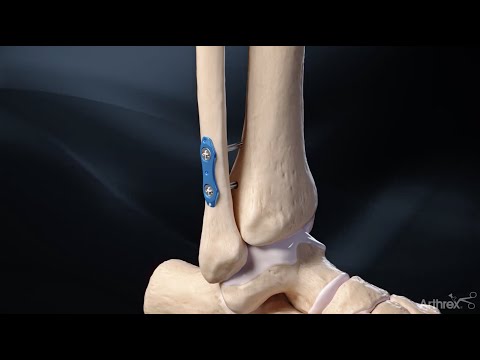
Why I use an Arthrex Syndesmotic TightRope?
Traditional screws rigidly hold the tibia and fibula together. They work, but they block the small natural motion of the syndesmosis and often require a second surgery for screw removal. These are used when the syndesmosis injury is associated with bad fractures and gross instability.
A Syndesmotic TightRope is a suture‑button construct that stabilizes the bones while permitting micro‑motion that more closely matches normal mechanics. In my hands, this helps with comfort during rehab and reduces the likelihood of routine hardware removal. In complex injuries or where added stability is needed, I may use two TightRopes or combine the device with plate fixation for an associated fibula fracture.
WHY SCOPE THE ANKLE?
High ankle sprains frequently come with inside‑the‑joint problems that X‑rays can’t show. Through tiny incisions, ankle arthroscopy lets me see the cartilage surfaces directly, remove loose fragments, and treat osteochondral lesions of the talus. Depending on size and location, I use techniques such as chondroplasty to smooth frayed cartilage and bone‑marrow stimulation (microfracture or drilling) to encourage healing in contained defects. Arthroscopy also allows me to assess the deltoid ligament on the medial side. If the deltoid is torn and the ankle remains unstable medially after syndesmotic reduction, I repair it using suture anchors, either arthroscopically or through a small open approach. Addressing these companion injuries at the same time as the syndesmosis repair reduces the risk of persistent pain or recurrent instability.
MY APPROACH
- Precise reduction
Under imaging, I realign the fibula and tibia to restore normal spacing and rotation. Small adjustments matter, because even a few millimeters can change contact pressures across the ankle.
- Arthroscopic Assessment
I inspect the joint for cartilage injury, remove loose bodies, debride inflamed tissue, and treat osteochondral lesions as indicated. I check the medial gutter for deltoid tearing. If instability persists medially after the syndesmosis is reduced, I perform a deltoid repair.
- TightRope fixation
I place one or two TightRopes across the syndesmosis to hold the reduction. The suture‑button construct is tensioned to restore anatomic alignment without over‑compression. If there is a related fibular fracture, I stabilize the fracture first and then address the syndesmosis.
- Final check under load
I test stability with gentle stress to confirm the ligaments and fixation are doing their job.
RECOVERY TIMELINE
Recovery is individualized, but a typical plan includes a short period of immobilization to let the soft tissues settle, followed by early, controlled range of motion. Weight bearing progresses in a boot when swelling and pain allow, often in the first few weeks for isolated syndesmotic repairs and slightly later if cartilage work or deltoid repair was required. Physical therapy focuses on restoring motion, calf strength, balance, and gait mechanics. Desk work may be possible within a few weeks. Jobs that require prolonged standing, ladder use, or carrying loads demand a more cautious ramp‑up. Athletes usually follow criteria‑based return protocols that include hopping, cutting, and sport‑specific drills once strength and balance normalize and swelling is minimal.
LONG TERM CONSEQUENCES?
The goal is a stable, painless ankle that tolerates daily life, work demands, and athletic loads. Most patients regain excellent function when instability is corrected and cartilage or deltoid injuries are treated at the same sitting. Some stiffness or swelling can linger for several months, especially after cartilage procedures. I monitor progress closely and adjust therapy based on milestones rather than the calendar.
- Workers’ compensation considerations
For work‑related injuries, the operative report, arthroscopy photos, and weight‑bearing imaging provide objective evidence of the problem and the repair. I translate those findings into clear restrictions that match real job tasks, which helps employers plan modified duty and reduces friction during the return‑to‑work process. If your claim involves denials around imaging or physical therapy, my team documents medical necessity and timelines so your attorney has what they need.
- Why patients choose my approach
I focus on precision diagnosis with weight‑bearing imaging, minimally invasive arthroscopy to treat what is actually wrong inside the joint, and flexible syndesmosis fixation that supports a smoother rehab. That combination is designed to protect cartilage, restore alignment, and get you back to work and sport as safely and efficiently as possible.
Frequently asked questions
- Is TightRope better than screws?
Both methods can work. I prefer TightRope fixation for most syndesmotic injuries because it allows small natural motions while maintaining stability and usually does not require hardware removal. In certain fracture patterns or severe instability, I may use supplemental fixation.
- Will I need a second surgery to remove hardware?
Suture‑button constructs typically stay in place. Screw fixation often requires removal, especially if it causes pain or limits motion. I discuss the plan with you based on your injury pattern and job demands.
- Do you always repair the deltoid ligament?
No. I repair the deltoid when it is torn and the ankle remains unstable medially after the syndesmosis is reduced. Treating true medial instability improves alignment and helps prevent ongoing pain.
- What happens if you find cartilage damage?
Small, stable lesions are smoothed; larger contained lesions often receive bone‑marrow stimulation. Very large or cystic lesions may need staged or graft procedures. The rehab plan accounts for any cartilage work performed.
- How soon can I walk after surgery?
Weight bearing depends on the specific repair. Many patients begin protected weight bearing in a boot within the first few weeks, advancing as pain, swelling, and strength allow. Jobs with heavy physical demands take longer to resume safely.
- Do you treat high ankle sprains from workplace injuries?
Yes. I regularly manage workers’ compensation ankle injuries in Pittsburgh and Monroeville and provide the documentation required for safe restrictions and return‑to‑work planning.
References
- https://www.arthrex.com/foot-ankle/knotless-tightrope-syndesmosis
- https://www.youtube.com/watch?v=rebFcIB_XvI
- Clanton TO, Viens NA, Campbell KJ, et al. Syndesmosis injuries in athletes. Journal of the American Academy of Orthopaedic Surgeons.
- Schepers T. Acute distal tibiofibular syndesmosis injury: a systematic review. Foot & Ankle International.
- Thornes B, Shannon F, Guiney AM, et al. Suture‑button versus screw fixation in the treatment of syndesmosis injuries: a randomized trial. Foot & Ankle International.
- Cottom JM, Hyer CF, Philbin TM, Berlet GC. Suture‑button fixation of the distal tibiofibular syndesmosis. Foot & Ankle Specialist.
- Shimozono Y, Hurley ET, Nguyen JT, et al. Microfracture for osteochondral lesions of the talus: a systematic review. Arthroscopy.
- Hintermann B, Regazzoni P, Lampert C, et al. Medial ankle instability and deltoid ligament repair. Foot & Ankle Clinics.
- American Orthopaedic Foot & Ankle Society. High ankle sprain (syndesmosis) patient resource.
- AAOS OrthoInfo. Sprained ankle patient education resource.
